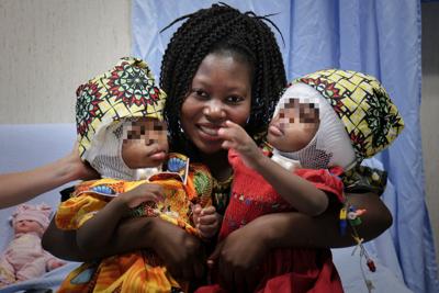
The headlines on RAI’s television news program on July 7, 2020 reported the successful operation of Siamese twins, joined at the head, at the Vatican’s Bambino Gesu Paediatric Hospital in Rome.
The case, of the two little girls from the Central African Republic, was studied for a year before the operation took place.
What is their mother Erminia’s dream now? That the girls “become doctors and give back to others what they received.” She would also like the girls to be baptized by Pope Francis.
This “extraordinary intervention” was prepared for over a year of study and in several surgical phases, specified in Italian a press release of the Hospital. The little girls, who arrived in Rome from Africa, were conjoined at the head, with “one of the rarest and most complex forms of cranial and cerebral fusion.” They “shared the bone of the posterior area of the cranium and the venous system. They are fine now.”
We translate the greater part of the press release, except for certain technical details. The remarks of specialists that point out our errors on the technical terms will be welcome.
“It’s the first case in Italy — and probably the only one in the world (similar operations aren’t described in the literature) — of a successful intervention on a pair of total posterior craniopagus, one of the rarest forms of fusion and the most complex at the cranial and cerebral level. Joined at the back of the head, they had the cranium in common and a large part of the venous system. More than a year of preparation and study, with the aid of advanced imagery systems and surgical simulation, ended in three very delicate interventions. The last, the definitive separation on June 5 of this year, was an 18-hour operation involving more than 30 persons, between doctors and nurses. A month later, the little girls are doing well. The have just celebrated their 2nd birthday and are hospitalized in the neurosurgical service sector of the Holy See’s Hospital, in two beds next to each other <and are> with their mother.”
The Pope’s Meeting at Bangui’s Hospital
“In July 2018, the President of the Vatican’s Hospital, Mariella Enoc, was on mission in Central Africa, in the capital Bangui, to follow the extension works of the paediatric facilities desired by Pope Francis. It was there that she met the newborn twins and decided to take them in hand, bringing them to Rome to give them a better chance of survival. . “When you meet with lives that can be saved, that must be done. We cannot and must not look the other way,” said President Enoc during a press conference on July 7.”
“Ervina and Prefina were born in the Medical Center of Mbaiki, a village 100 kilometers from Bangui. Their mother, Erminia, and the doctors only discovered that they were Siamese twins at the moment of the delivery by cesarean. However, the small health center was not equipped to handle the case, so the family was taken to the Central African capital.”
Arrival at Rome’s “Bambino Gesu”
“The mother and twins arrived in Italy on September 10, 2018 in the framework of the international humanitarian activities of the Holy See’s Paediatric Hospital. After some months at the Bambino Gesu of Palidoro, where they began a course of neuro re-education, the children were transferred to the neurosurgical service of the Janiculum for studies on the feasibility of the separation procedures. The first studies confirmed that the twins were in general good health; the neurological and clinical parameters were normal. However, there was a difference of arterial pressure: the heart of one of the little girls worked harder to maintain the physiological balance of the organs of the two, including the brain.”
Conjoined but Different
“Ervina and Prefina were joined in the parietal and occipital region of the cranium., that is, a large surface at the back of the head which includes the nape of the neck. They had cranial bones and skin in common; at a deeper level, they shared the <sickle> and the tentorium (fibrous membranes that separate the two cerebral hemispheres and those of the cerebellum) as well as a large part of the venous system (the network of vessels in charge of transporting the blood used by the brain to the heart to be re-oxygenated) which represented the most difficult challenge, for the neurosurgical team of the Bambino Gesu, in planning the interventions. From the fact of this particular conformation, the little girls entered the very rare category of “total” craniopagus twins, joined, that is, at the cranial and cerebral level. They had many things in common, except for different and distinct personalities: Prefina <is> playful and animated, Ervina more serious and attentive. A system of mirrors was used in the framework of the process of re-education to make them <get to> know each other and recognize each other, also by visual contact, before the separation.”
A Study that Lasted Over a Year
“The case of Ervina and Prefina was very difficult. To have them survive separately, all aspects had to be studied, planned down to the least detail. With this objective, a multi-disciplinary group was formed, made up of neurosurgeons, anesthetists, neuroradiologists, plastic surgeons, neuro re-educators, engineers, nurses in different specialized domains and physiotherapists.
The Ethics Committee was involved and shared a therapeutic course that could give the two girls the same chances of quality of life. The Bambino Gesu team developed the program on the basis of experience acquired with previous cases of successfully separated Siamese twins. Over the month, the twins were also prepared for the separation: with neuro re-education, they attained a level of cognitive and motor development similar to that of their peers. With the aid of numerous postural systems, which helped them pass their days in the best position possible, they faced the complex phases of the treatment. Thanks to a system of mirrors, they learned to recognize each other’s face and expressions and to establish a visual relationship. “
“Before passing to the surgical phases, the complex case of the Bangui twins was also presented and discussed at the international level, at New Delhi in India, where the first world conference was held of Siamese twins surgery in February 2019. In the Hospital’s history, it was the fourth case of separation of Siamese twins: in 2017, Algerian twins were joined at the chest and abdomen (thoraco-omphalopagus twins) and little Burundians joined at the sacrum (pygopagus twins). The first operation of this type took place in the 80s on two boys joined at the chest and abdomen.”
The Three Stages of Separation
“The great challenge for the success of the separation was the cerebral venous — the network of blood vessels (sinus veineux) system that the twins shared in several places. The surgery of the venous structures of the brain is complex and the risk of bleeding and ischemia is high the Bambino Gesus’s neurosurgical team decided to proceed by stages: three very delicate interventions to reconstruct progressively two independent venous systems capable of containing the flow of blood that circulates from the brain to the heart.”
“The first intervention: in May 2019 the twins entered the operating room to begin to fashion new autonomous venous structures: the neurosurgeons separated a part of the tentorium and the first of two common transversal sinuses that will affect each of the girls; then, with bio-compatible materials, they reconstructed a membrane capable of maintaining the divided cerebral structure before the definitive separation.”
“The second intervention was in June 2019. The team, assisted by an anesthetist group separated the superior sagittal sinuses (the posterior half of the venous channels that extend between the two cerebral hemispheres) and the point of junction of the venous sinuses of the brain (. . . ). It was a crucial phase: the operational space is of some millimeters and the neurosurgeons proceeded under the direction of a neuro-navigator. “
“A year later, June 5, 2020, was the moment of the definitive separation. The little girls had grown, the new venous architecture was consolidated and functioning; the portion of skin necessary to cover the cranium of each of the little ones was enlarged with “expanders,” positioned some months earlier with a series of plastic surgery operations and <then> the last phase could be launched. A team of over 30 persons was ready in the operating room, including doctors, surgeons and nurses. The operation lasted 18 hours: first the skin “expanders” were removed, then the second transversal sinus and its tentorium were separated; finally, the bones of the cranium that kept the two little girls together were divided. Once the twins were separated, the operation continued in two different operating rooms, with two distinct teams, to reconstruct the membrane that covers the brain, remodel the cranium’s bones and recreate the mucous skin.”
“It was an exciting moment, a fantastic and unforgettable experience,” said Carlo Marras, Head of Neurosurgery at the Bambino Gesu, and of the team that cared for the twins. “It was a very ambitious objective and we did everything possible to attain it, with passion, optimism and joy, sharing each stage and studying each detail together.”
The Role of Technology: 3-D Reconstruction and Neuro-navigator.
“Each phase of the twins’ itinerary was studied and planned with the help of advanced imagery systems available at the Hospital: computed tomography and three-dimensional magnetic resonances, 4D angiography, 3D reconstruction software, neuro-simulator. Thanks to these technologies, combined with one another, the little girls’ cranium was recreated in 3D with all the internal anatomic details, including the vascular network. As things progressed, it was possible to evaluate the functionality of the individual cerebral structures, to quantify the blood flow and to make a prediction on the functioning of the new system after the interventions. The most advanced neuro-navigation systems were used in the operating room. Particularly useful tools, in such complex and rare cases, which point out to the surgeon, with millimetric precision, the position of the most delicate structures.”
Ervina’s and Prefina’s Future
“A month after the definitive separation, the twins are doing well. <After> a few follow-up days in intensive care, they returned to their room, a room with two simple beds. On June 29 they celebrated their second birthday, looking at each other in the eyes, moving their small hands to the rhythm of the music, in their mother’s arms. They went through very difficult operations; the wounds will take time to heal; the risk of infection is always present. The neuro re-education program continues and in a few months they will have to wear a protective helmet. However, the post-operational controls indicate that the brain <in each one> is intact. The recreated system is functioning, the blood flow has adapted to the new way. The doctors of the Neuro-Sciences Department explained that <the girls> are in a state that will give them the opportunity to grow normally both from the motor as well as the cognitive point of view, and to lead a normal life, as all girls of their age.”
“Moved, after today’s press conference, their mother, Erminia, thanked the Hospital and all the persons that took care of her children: “Ervina and Prefina were born twice. If we had stayed in Africa, I don’t know what fate they would have had. Now that they are separated and doing well, I would like them to be baptized by Pope Francis, who has always taken care of the children of Bangui. My little ones can now grow, study and become doctors to save other children.”
Total Posterior Craniopagus Is Very Rare
The birth of Siamese twins is a very rare event and, among the different types, the twins joined at the head (craniopagus) are the rarest: one case in 2.5 million live births, five cases out of 100,000 twins, in particular for girls. In the scientific literature, only some ten cases are described. Craniopagus is defined as “partial” when the contact point between the two heads is limited to bones and skin; “total” when the fusion includes the cerebral structures and, in particular, the venous system. There are also differences among total craniopagus: the most “common” are twins joined at the top of the head <vertical craniopagus), rarer are those joined at the nape of the neck (posterior craniopagus).”
“According to the available data a few years ago, 40% of craniopagus <twins> were dead at birth. For the remaining 60%, the hope of life has not surpassed 10 years. Up to the 60s, the attempts at separation of total craniopagus <cases> had a mortality rate close to 100%. Thereafter, with the development of technology and the introduction of phased surgery, the survival, the attempts and the quality of life have increased. In the course of the last 20 years, there have been in Europe two total craniopagus <twins> separated successfully: they are two pairs of twins joined at the top of the head (vertical) operated on in several stages in London. No case is described in the literature, however, with the characteristics of the Bangui twins, with total craniopagus joined at the base of the neck (posterior).”